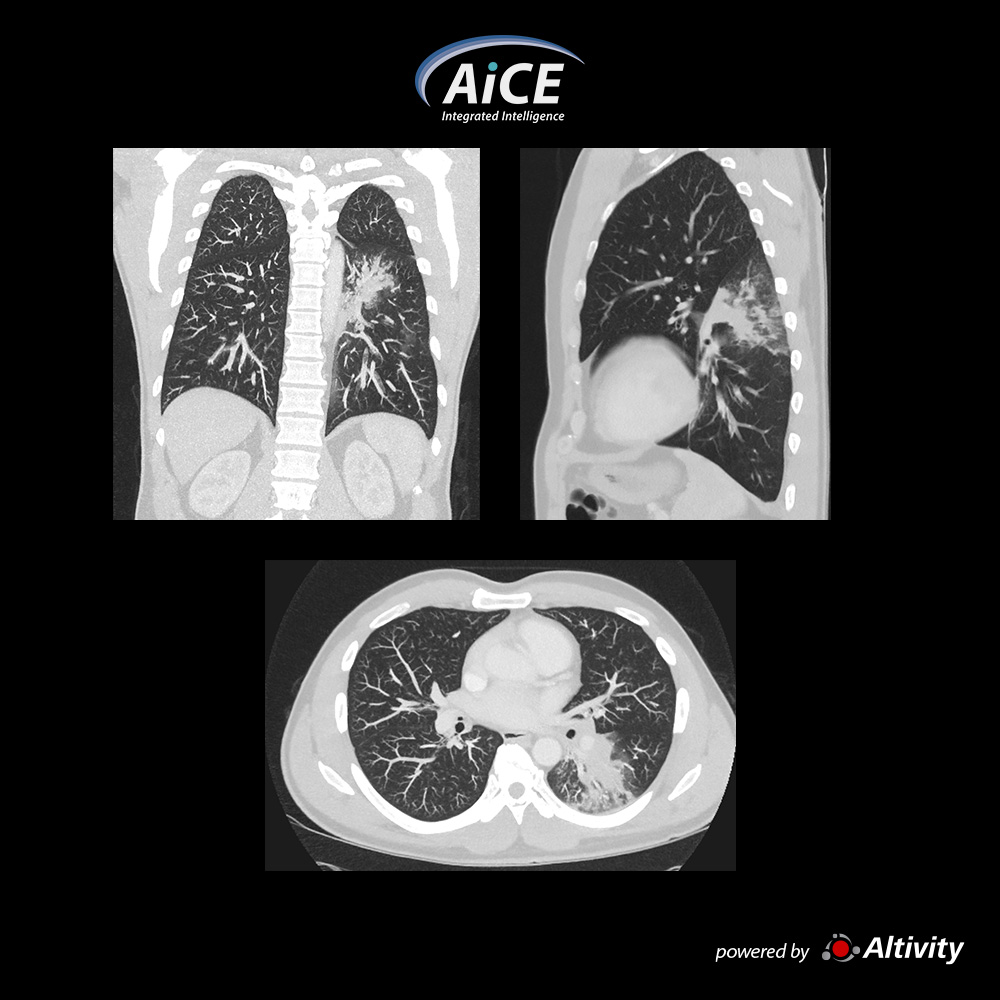
A pneumonia observada nas imagens de MPR do homem de 49 anos com dispneia e IMC de 25,4
Cortesia de South Coast Radiology, Qld, Austrália
View Scan Parameters
Modo de varreduraUltra helicoidal
Colimação0.5 mm × 80
kVp120
mAsSURE Exposure
Tempo de rotação0.35 s
Faixa de varredura409 mm
Redução de DoseAiCE
CTDIvol4.5 mGy
DLP204.4 mGy·cm
Dose efetiva2.9 mSv
Fator k0.014 (AAPM Report 96)
×
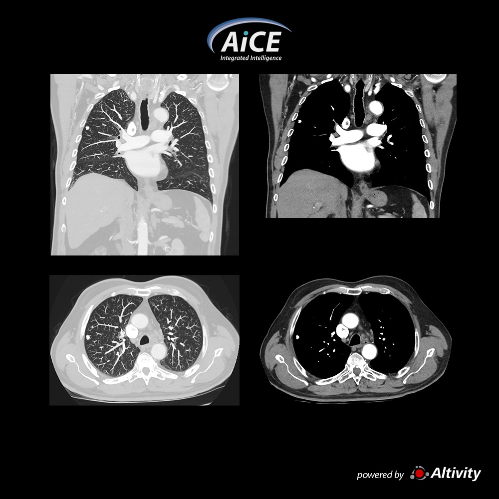
Tórax – nódulo calcificado
Homem de 82 anos, a tomografia do tórax de acompanhamento demonstra um nódulo calcificado no pulmão direito
Cortesia de South Coast Radiology, Qld, Austrália
View Scan Parameters
Modo de varreduraUltra helicoidal
Colimação0.5 mm × 80
kVp120
mAsSURE Exposure
Tempo de rotação0.35 s
Faixa de varredura364.5 mm
Redução de DoseAiCE
CTDIvol5.2 mGy
DLP214 mGy·cm
Dose efetiva3.0 mSv
Fator k0.014 (AAPM Report 96)
×
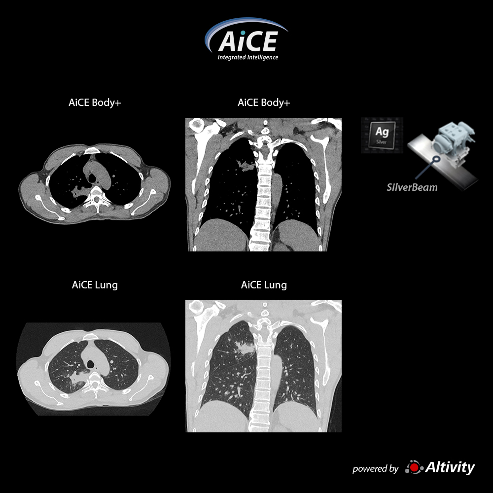
Low dose chest with SilverBeam
SilverBeam in combination with AiCE Deep Learning Reconstruction provides excellent image quality at dose levels more comparable to a chest x-ray.
In this patient the DLP was only 24 mGy.cm with an effective dose of 0.3 mSv.
Courtesy
Medic Medical Center, HCMC Vietnam
View Scan Parameters
Scan ModeSilverBeam Ultra-Helical
Collimation0.5 mm × 80
kVp120
mAsSUREExposure
Rotation Time0.5 s
Scan Range241 mm
Dose ReductionAiCE
CTDIvol0.70 mGy
DLP24.70 mGy-cm
Effective Dose0.34 mSv
k-factor0.014 (AAPM Report 96)
×
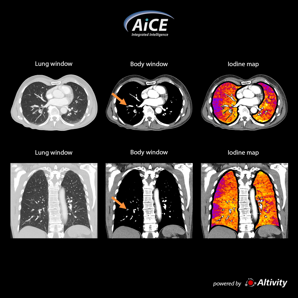
Pulmonary CTA - Iodine Mapping SCT
Color iodine maps quickly and definitively show areas of hypoperfusion to enhance your diagnosis.
Hyperperfused areas are commonly associated with pulmonary pathology as demonstrated in this case of a pulmonary embolism.
Courtesy
Royal Bournemouth Hospital, Bournemouth, UK
View Scan Parameters
Scan ModeUltra-Helical
Collimation0.5 mm × 80
kVp120
mAsSUREExposure
Rotation Time0.35 s
Scan Range300 mm
Dose ReductionAiCE
CTDIvol1.6, 2.0 mGy
DLP126.9 mGy-cm
Effective Dose1.78 mSv
k-factor0.014 (AAPM Report 96)
×
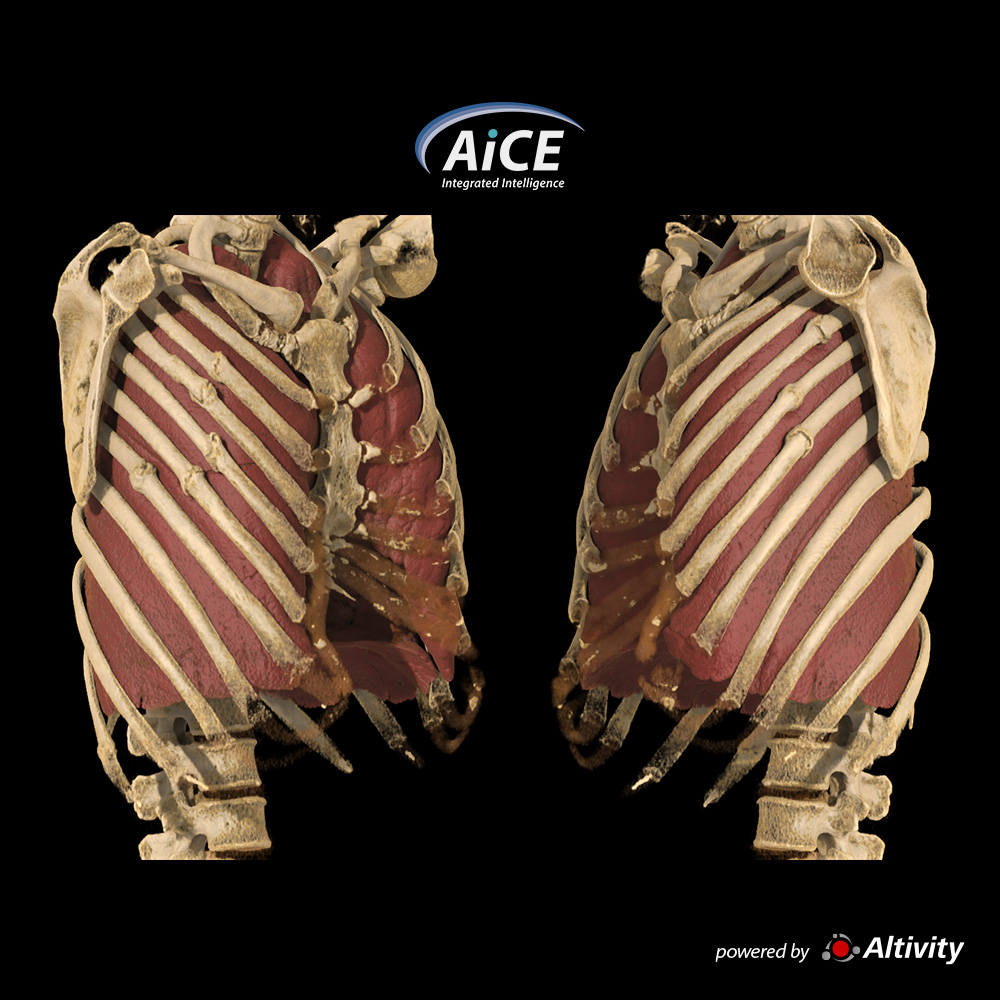
Fraturas de costelas
Mulher de 51 anos com histórico de queda anterior. A renderização do Global Illumination fornece excelente representação das várias fraturas das costelas
Cortesia de South Coast Radiology, Qld, Austrália
View Scan Parameters
Modo de varreduraUltra helicoidal
Colimação0.5 mm × 80
kVp120
mAsSURE Exposure
Tempo de rotação0.35 s
Faixa de varredura376 mm
Redução de DoseAiCE
CTDIvol2.9 mGy
DLP123.5 mGy·cm
Dose efetiva1.7 mSv
Fator k0.014 (AAPM Report 96)
×
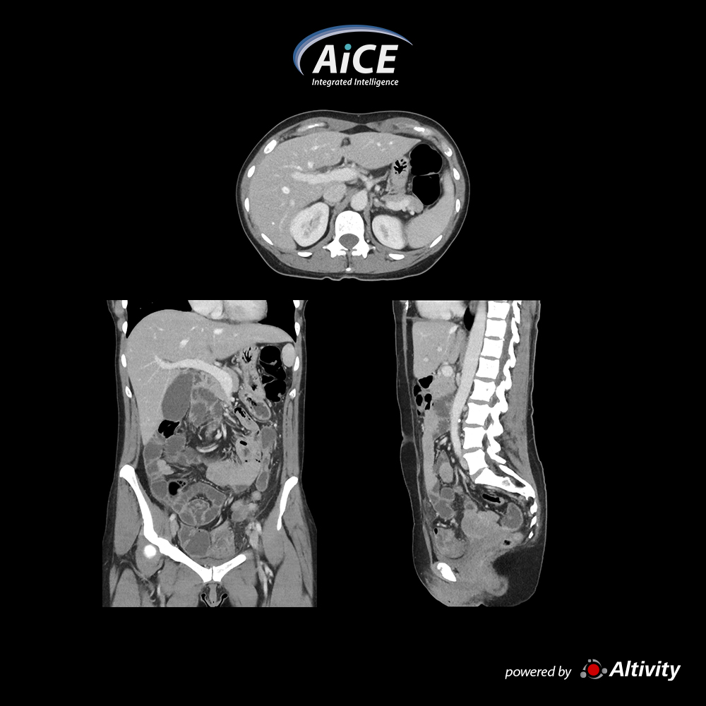
Portal Abdomen
Low noise, excellent image quality in this portal phase abdomen scan
Courtesy
Royal Bournemouth Hospital, Bournemouth, UK
View Scan Parameters
Scan ModeUltra-Helical
Collimation0.5 mm × 80
kVp120
mAsSUREExposure
Rotation Time0.5 s
Scan Range449 mm
Dose ReductionAiCE
CTDIvol4.3 mGy
DLP213.9 mGy-cm
Effective Dose3.2 mSv
k-factor0.015 (AAPM Report 96)
×
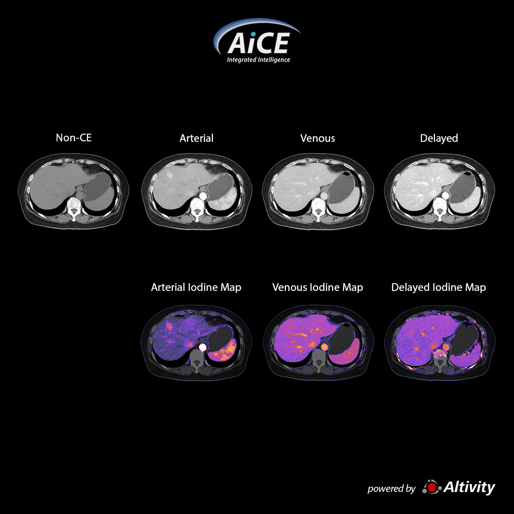
Multi Phase Abdomen – Iodine Mapping SCT
Color iodine maps can be provided with every multiphase abdomen protocol to further enhance your diagnostic confidence.
Iodine mapping can help to visualize and differentiate hypervascular lesions such as hepatocellular carcinoma (HCC) as seen in this case.
Here a hyper-vascularized arterial lesion can be easily visualized in both the gray scale image and color iodine maps. The lesion also demonstrates a “washout” pattern in the venous and delayed phase consistent with imaging findings associated with HCC.
Courtesy
Medic Medical Center, HCMC Vietnam
View Scan Parameters
Scan ModeUltra-Helical
Collimation0.5 mm × 80
kVp120
mAsSUREExposure
Rotation Time0.5 s
Scan Range456, 456, 456, 200 mm
Dose ReductionAiCE
CTDIvol4.8, 4.8, 4.8, 4.8 mGy
DLP833 mGy-cm
Effective Dose12.49 mSv
k-factor0.015 (AAPM Report 96)
×
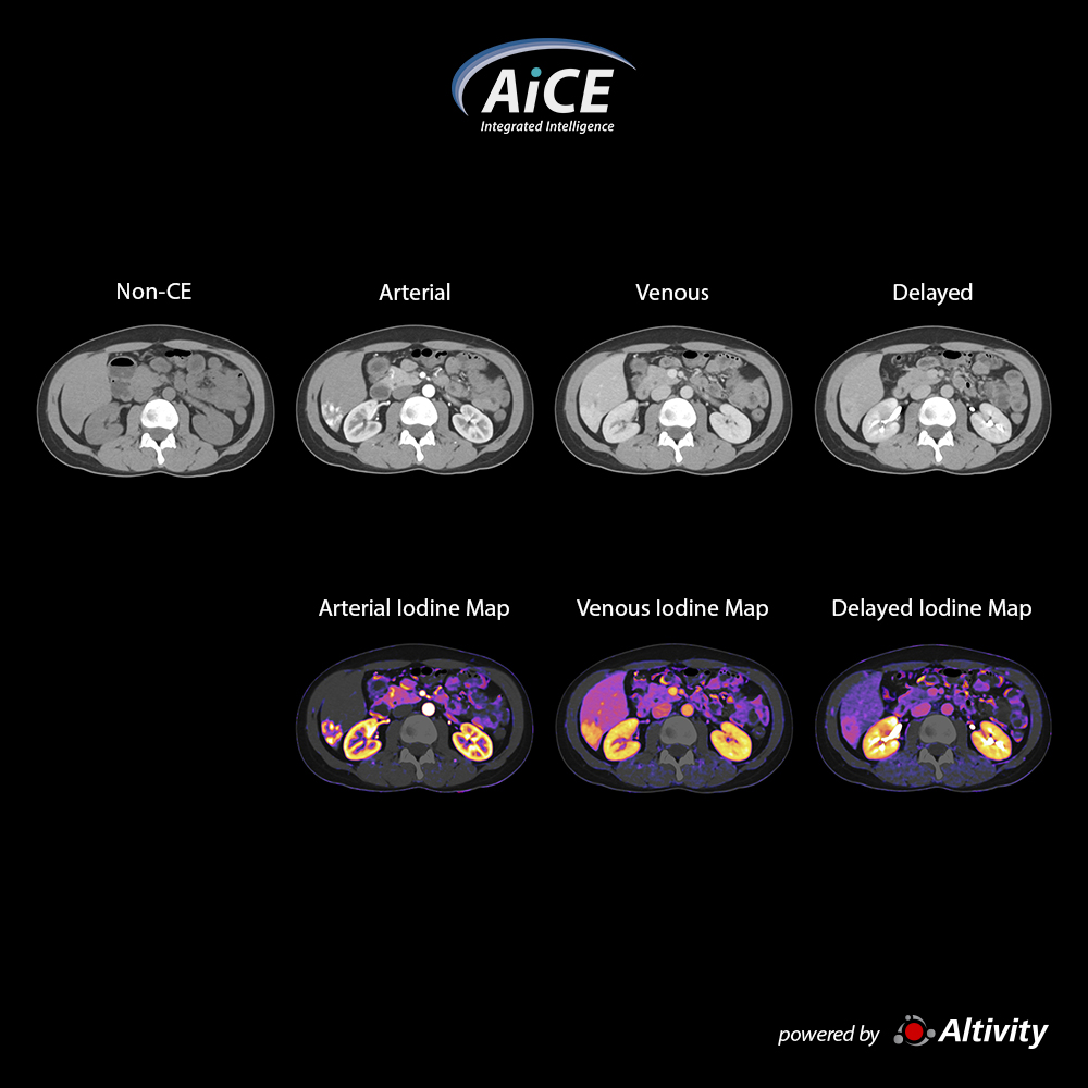
Multi Phase Abdomen – Iodine Mapping SCT
Iodine mapping can help to visualize and differentiate hypervascular lesions that may exhibit similar enhancements in the arterial phase.
Here a hyper-vascularized arterial lesion can be easily visualized in both the gray scale image and color iodine maps.
The iodine maps unequivocally show the lesion still retains some contrast material in the venous and delayed, but the enhancement is less intense compared to the arterial phase.
Courtesy
Medic Medical Center, HCMC Vietnam
View Scan Parameters
Scan ModeUltra-Helical
Collimation0.5 mm × 80
kVp120
mAsSUREExposure
Rotation Time0.5 s
Scan Range288, 280, 452, 333 mm
Dose ReductionAiCE
CTDIvol4.8, 4.8, 4.8, 4.8 mGy
DLP876 mGy-cm
Effective Dose13.14 mSv
k-factor0.015 (AAPM Report 96)
×
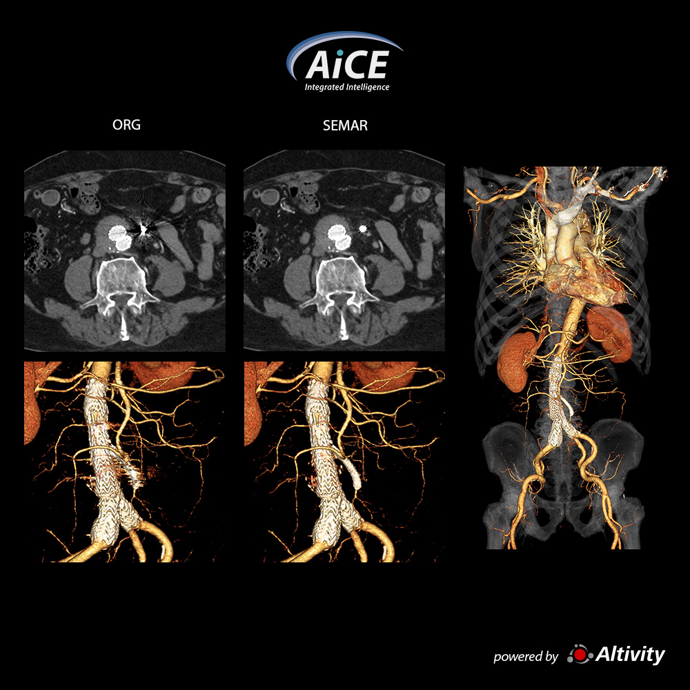
Aorta CTA + SEMAR
O SEMAR remove os artefatos do enxerto de stent na CTA da aorta abdominal
Cortesia de Hakujyuji Hospital, Japão
View Scan Parameters
Modo de varreduraUltra helicoidal
Colimação0.5 mm × 80
kVp120
mAsSURE Exposure
Tempo de rotação0.5 s
Redução de DoseAiCE
CTDIvol7.9 mGy
DLP550.3 mGy·cm
Dose efetiva8 mSv
Fator k0.0145 (AAPM Report 96)
×
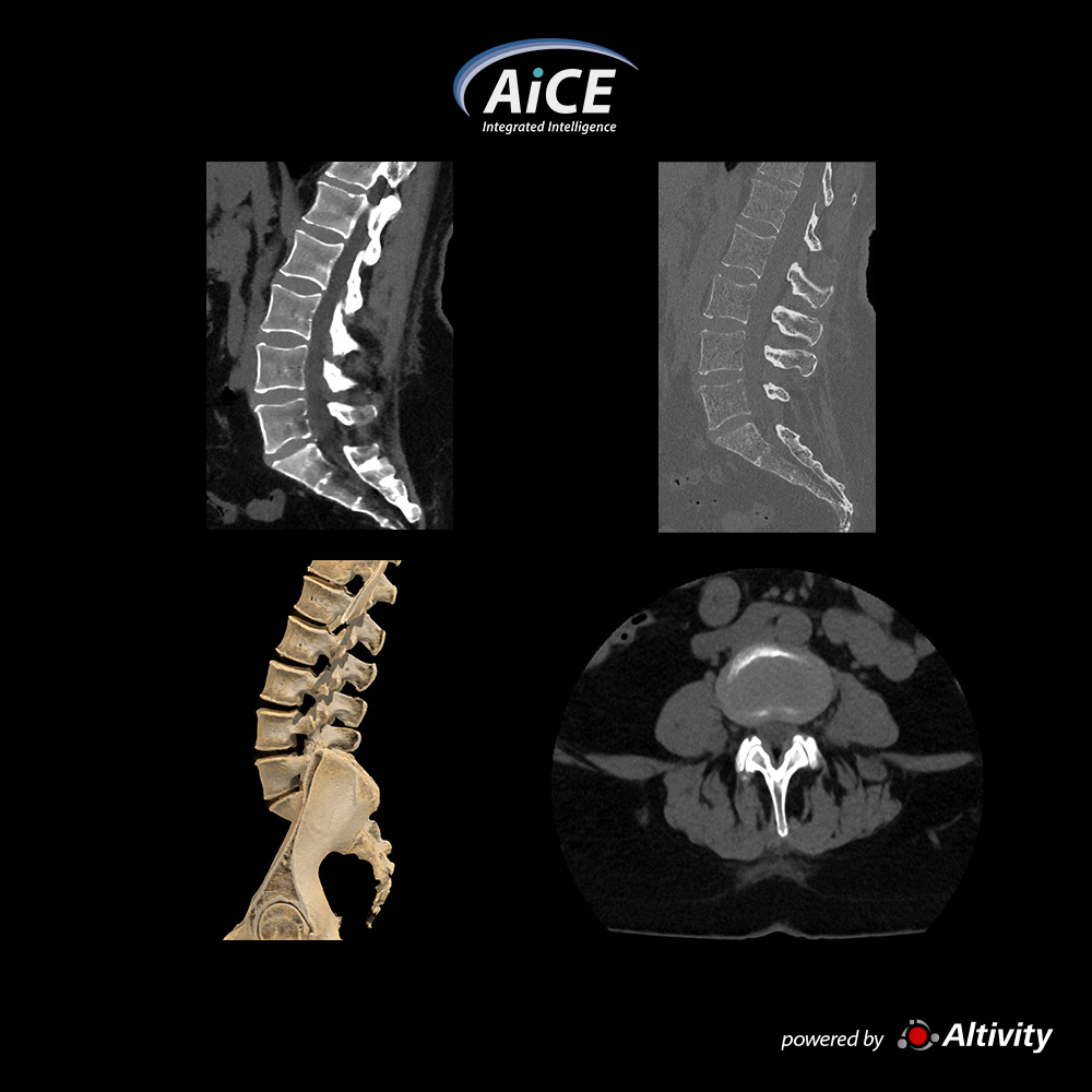
Coluna Lombar
Reconstruções de alta qualidade do tecido mole e osso com o AiCE
Cortesia de South Coast Radiology, Qld, Austrália