Imaging | Computed Tomography
Our CT innovations have been designed for accurate and comfortable oncology imaging with simplified workflows, high spatial resolution and automated dose reduction.

Aquilion Precision
Precision in Every Detail
Introducing the world’s first Ultra-High Resolution CT (UHR CT), capable of resolving anatomy as small as 15 microns while delivering the image resolution of a catheterization laboratory.
Ultra-High Resolution CT

1024 x 1024 Matrix Aquilion Precision UHR CT
New Technologies:
- UHR Detector
- UHR DAS
- UHR Tube
- UHR Gantry
- UHR Reconstruction
0.25 mm
Detector
150 micron
Spatial Resolution
50 lp/cm at 0% MTF
Reference only
Whitepaper
Aquilion Precision Ultra-High Resolution CT: Quantifying diagnostic image quality
Kirsten Boedeker, PhD, DABR Senior Manager, Quantitative Image Quality, Canon Medical Systems Corporation
New UHR Reconstruction Matrix
0.5 mm
896 channel
Detector


0.25 mm
1792 channel
UHR Detector


Clinical Example
Lung1
SHR / 0.25 mm
- Courtesy of Kyorin University, Japan


Lung2
HR / 0.5 mm
- Courtesy of Prof. Prokop RadboudUMC, Nijmegen, the Netherlands


PUREViSION Optics
Aquilion Prime SP strikes the perfect balance between image quality and dose – transforming routine imaging with new levels of detail, resolution and safety.

Whitepaper
PUREViSION Optics: The optimal balance between image quality and dose
Jay Vaishnav, PhD Clinical Scientist, Manager, Medical Affairs, Canon Medical Systems USA, Inc.

vHP3*—Variable Helical Parameters
Overall faster scan times make vHP3 compliant to the needs of the patient, and shorten exam times for trauma imaging when every second is critical.
With this technology, a single series reconstruction enables several studies to be interpreted simultaneously for faster scans when every second counts.
vHP3 may also help you:
- Decrease radiation dose
- Decrease the amount of IV contrast needed
- Improve the workflow of complex clinical exams
- Option
Easy Patient Positioning
Aquilion Prime SP’s accessible gantry and couch have been designed for enhanced comfort, particularly for bariatric patients or those who are elderly and living with cancer.
- Option

Aquilion Prime SP's accessible gantry design and powerful advanced imaging applications deliver an ideal oncology solution to assist clinicians with visualization, staging and treatment planning.

Adaptive Iterative Dose Reduction 3D

Single Energy Metal Artifact Reduction (SEMAR)

SURESubtraction unique Differential Iodine Enhancement

Aquilion ONE / GENESIS Edition
Transforming CT and Oncology care
Clinically-focused research and technological developments have culminated in a CT system that reduces dose and simplifies workflows for tumor visualization and assessment.
System Highlights:
- Up to 16 cm z-axis coverage
- 0.5 mm × 320 PUREViSION detector row
- 0.275 sec rotation
Tumor Assessment with Whole Organ Perfusion
Volume Perfusion Liver*
With the ability to evaluate tumor response or anti-angiogenesis, CT body perfusion has become a valuable clinical tool – especially now that you’ve got 16 cm of z-axis coverage at your disposal.
Liver Perfusion Protocol: DLP = 720.8 mGy.cm, Effective dose = 10.81 mSv (k = 0.015)
- CT Volume perfusion only available with Aquilion ONE

Outstanding Lesion Visualization and Assessment
SURESubtraction Body
Clinical indication:
- Assessment of tumors, lymph nodes and vascular lesions
- Infarcts, ischemia
Iodine mapping:
- Blood flow mapping
- Visualize local perfusion differences
- Display local iodine concentration
- Visualize contrast enhancement
Differential enhancement maps:
- “Pure” arterial and venous maps

Expert Tools
- CT Volume perfusion only available with Aquilion ONE


Learn More Product Details
Imaging | PET/CT
Celestion PUREViSION Edition
Confident Diagnosis with Lesion Metabolic Assessment
PET
- 88 cm PET bore
- 394 ps typical Time-of-Flight
- 19.6 cm axial field-of-view
- 4 mm x 4 mm crystals
CT
- 90 cm CT bore
- 70 cm true FOV / 85 cm EFOV
- PUREViSION 0.5mm x 16-row detector
- 32 mm coverage
- 32 slice coneXact reconstruction
- AIDR 3D dose reduction
- SEMAR, Metallic artifact reduction
The Largest PET/CT Bore and FOV
Designed to Help You See More
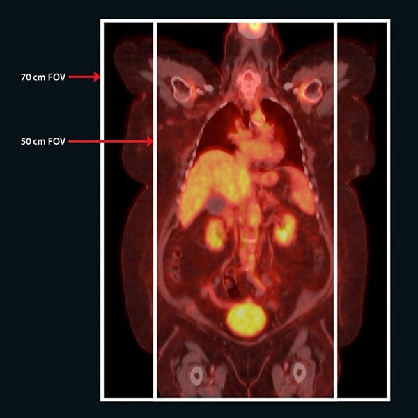
70 cm True Field of View
With oncology patients, accuracy is everything. To meet this need, Celesteion offers a true 70 cm PET and CT field of view (FOV).Overcome the challenge of a small 50 cm FOV to improve image quality and treatment planning.
90 cm Wide CT Bore
Celesteion’s wide bore is built for maximum patient positioning, comfort and peace of mind. The industry-leading 90 cm CT bore and 88 cm PET bore enhances your flexibility and workflow efficiency.Enjoy greater flexibility and efficiency with a class-leading bore that optimizes patient comfort.
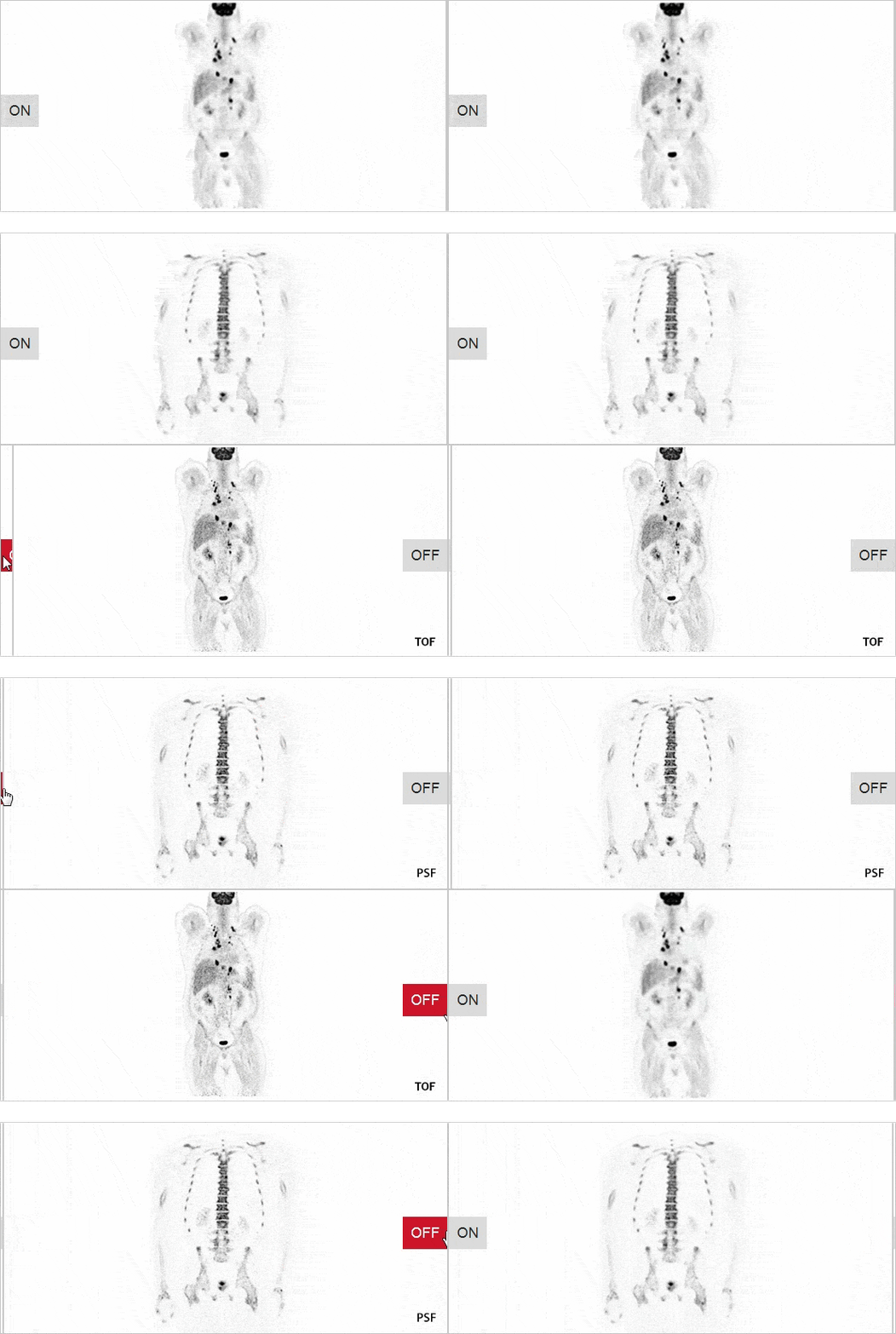
Time of flight (TOF) and point spread function (PSF) technology for greater accuracy
SUREFLiGHT Reconstruction Technology
Celesteion PUREViSION edition with TOF and PSF reconstruction can improve image resolution and possibly allow better lesion detection and follow-up.
- 394 ps typical TOF
- PSF reconstruction
- Optimal speed and image resolution
- Improved lesion detection
- Improved lesion localization
Learn More Product Details
Imaging | Magnetic Resonance
Our MRI solutions provide optimized workflow and image quality to help you deliver efficient and effective care to each of your oncology patients.

Unique Acoustic Noise Reduction Suites
Pianissimo Zen1

- 99% reduction by unit of Loudness level "dB" and 97% reduction by unit of perceived loudness "Sone"
- ¹ Optional

Whole body image with WFS DIXON
WFS* DIXON achieves consistent fat suppression and homogeneity while acquiring four different tissue contrasts in one scan, reducing the total number of scans needed.
Available for T1, T2, and PD sequences, WFS DIXON can be acquired in any area of the body.
- WFS: Water Fat Separation

Post-processing & 3D Visualization
Tumor Assessment
Our image processing software solutions help you view and analyze a range of tumor characteristics, including volume, cellularity (DWI), aggressiveness (DCE), and perfusion (DSC/IVIM).

- Optional, designed and manufactured by Olea Medical


Whitepapers
Women's Health:
Borderline Ovary Tumor(Borderline serous cystadenoma)
Men's Health:
Prostate Adenocarcinoma


Learn More Product Details
Imaging | Ultrasound
Canon Medical Systems’ comprehensive Aplio i-series / Prism Edition provides unprecedented resolution and penetration for quick and reliable diagnoses.

Comprehensive Experts Tools
- Availability dependent on system


Accurate Pre And Post-therapy Cardiac Function Assessment
2D and 3D* Cardiac Wall Motion Tracking
Evaluate cardiac function during/before treatment. This appears to be increasingly important prior, during, and after cancer treatments to monitor patients' overall cardiac health status.
Aplio’s advanced Wall Motion Tracking technology provides immediate visual and quantitative access to global and regional myocardial wall motion dynamics (in 2D and 3D) on both live and previously stored images.
- Availability dependent on system
Learn More Product Details

Social Media