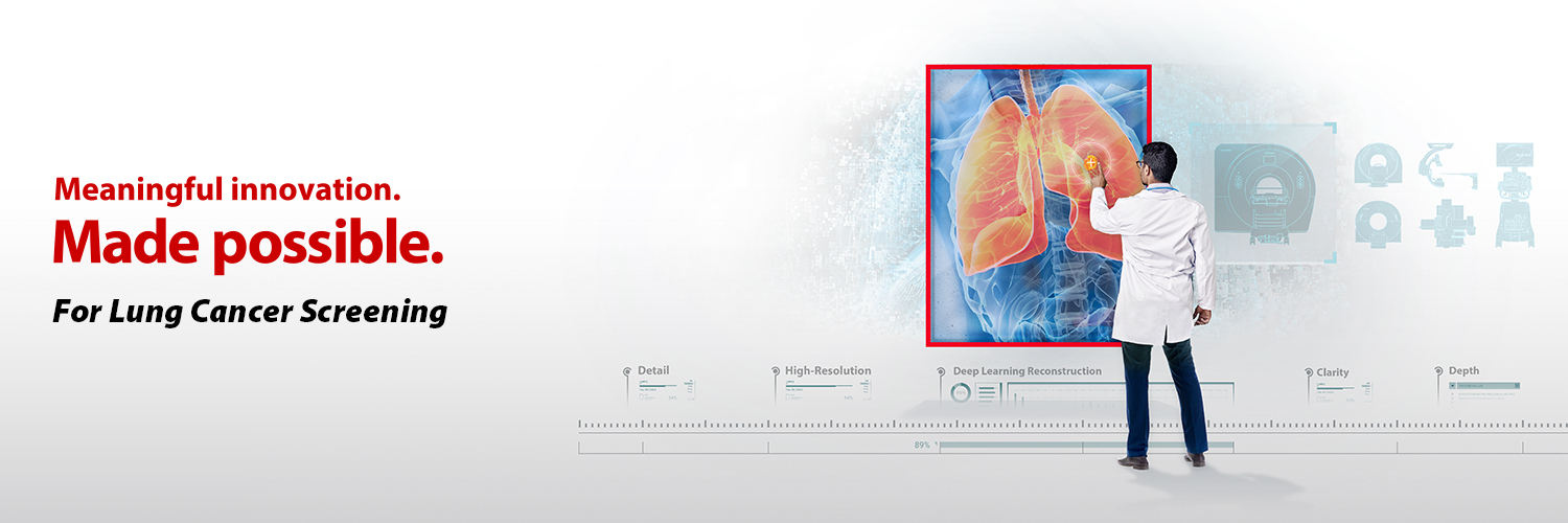
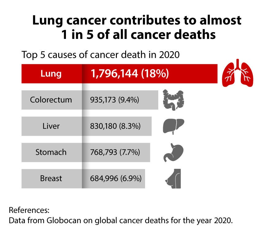
Through our "Screen for Life. Breathe with Hope." initiative, Canon Medical aims to change societal perceptions of lung cancer and encourage participation in early screening, which could ultimately contribute to the reduction of cancer mortality.2
Stay at the forefront of lung cancer screening insights from Canon Medical and top experts.
Subscribe now (Link to Canon Medical global website)
Expert Workshops at RSNA 2024
Missed the hands-on experience at RSNA?
On-demand video is coming soon. Subscribe now to access the full video and stay updated on future workshops.

Ultra-High Resolution Lung CT:
Pushing the Boundaries of Lung Imaging
Pushing the Boundaries of Lung Imaging
Russell Bull, MD
Consultant Radiologist
Royal Bournemouth Hospital
Bournemouth, UK
Consultant Radiologist
Royal Bournemouth Hospital
Bournemouth, UK

Diagnosis and Management of Lung Nodules in a Screening Setting
Mathias Prokop, MD, PhD
Professor of Radiology & Chairman of Department of Medical Imaging, Radboud University Medical Center Nijmegen, the Netherlands
Professor of Radiology & Chairman of Department of Medical Imaging, Radboud University Medical Center Nijmegen, the Netherlands
Latest insights
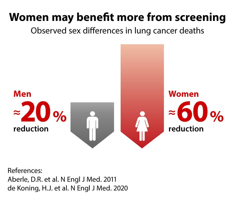
Lung cancer screening (LCS) saves lives, but more people need to get screened.
Get the big picture on challenges faced in screening and practical solutions in just one glance.
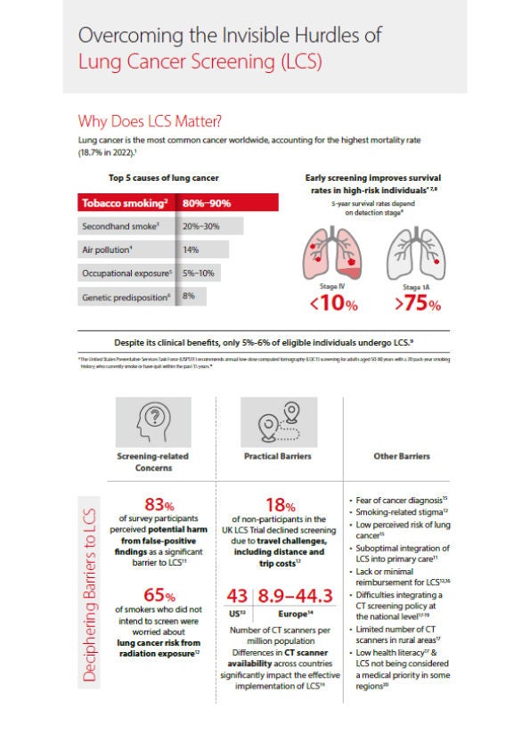
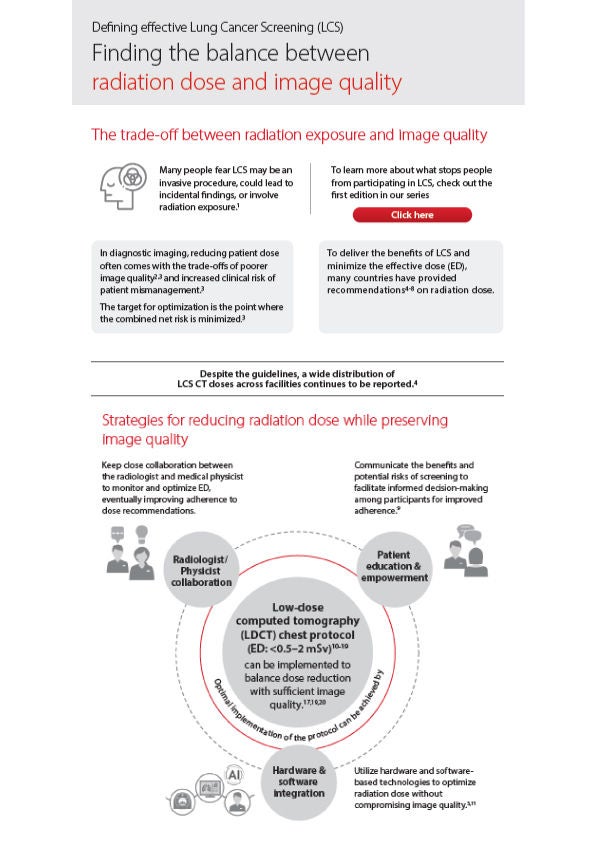
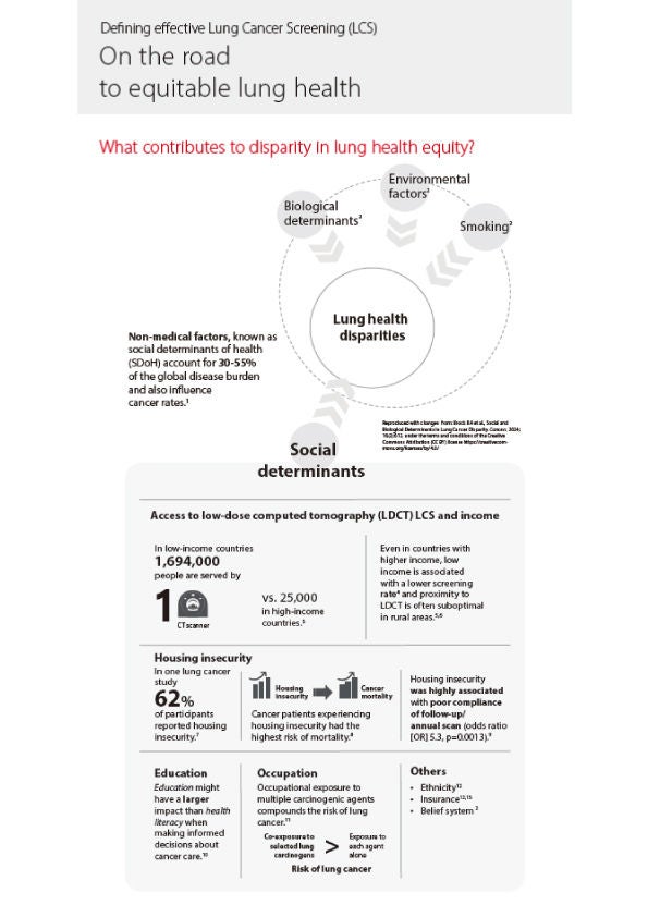
Subscribe to our newsletter to receive updates on our educational events and scientific information.
Subscribe now (Link to Canon Medical global website)
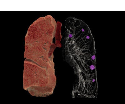
Lung cancer screening
Early detection to save more lives
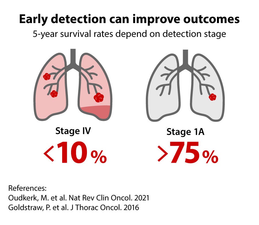
Lung cancer is one of the most frequent causes of cancer and the leading cause of cancer death globally.1 When patients present with symptoms, the disease often has already progressed, with limited options remaining for treatment. As a result, prognosis in lung cancer tends to be poor, despite advances in treatment. However, when the disease is detected at an early stage, the chances of survival increase dramatically.2
Consequently, there is a growing interest in lung cancer screening worldwide as the main opportunity to reduce lung cancer mortality.
Consequently, there is a growing interest in lung cancer screening worldwide as the main opportunity to reduce lung cancer mortality.
Canon Medical aims to be your partner in the fight against lung cancer and offers a unique set of lung cancer screening solutions. These include a suite of innovative technologies to enhance your clinical confidence everywhere, delivering high-quality imaging at low dose, streamlining workflows, and offering comprehensive diagnosis throughout the entire cycle of care.
Current challenges
Lung cancer screening faces various challenges that hinder its widespread success.
As part of our Made for Life philosophy, Canon Medical is committed to providing support to address these challenges.
Challenge 1: Low participation rate
Many individuals at high risk for lung cancer are often unaware of the potential benefits of screening or may face barriers such as cost, accessibility, or fear of the screening process.
Challenge 2: Workload and workflow
Workflow issues, such as managing the influx of screening participants and ensuring timely communication of results, can be a significant burden. Moreover, the repetitive nature of interpreting numerous lung scans can be mentally draining and lead to a loss of enthusiasm.
Challenge 3: Maintain clinical confidence at low dose
Screening participants are typically exposed to repeated CT scans which raises concerns around radiation dose. Lung cancer screening when performed without efficient CT hardware and image reconstruction algorithms, may yield inconclusive or false positive results, leading to additional and costly follow-up procedures, while causing anxiety and stress for patients.
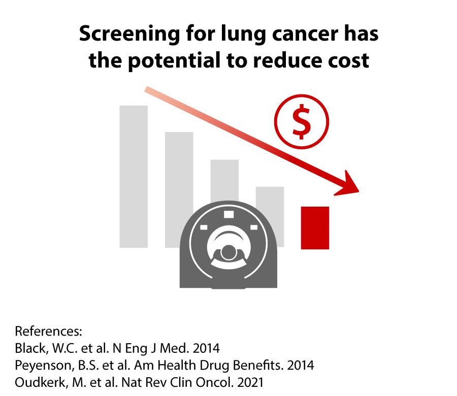
Challenge 4: Cost-effectiveness
Cost-effectiveness of the program requires a careful balance of the investments in equipment and staff, training, and the number of diagnostic and downstream follow-up procedures versus the benefits of improved health outcomes and less spending on costly therapies in the long-run.
Our solutions
We are taking measures to increase the accessibility of lung cancer screening for the public, providing high-quality imaging at low dose, removing workflow inefficiencies, supporting the interpretation and communication of findings among clinicians, and streamlining follow-up procedures.
Together, we can improve the patient experience and ensure a high-quality and cost-effective screening program, while minimizing the burden of the lung cancer on patients worldwide.


Easy access to screening
Easy access from anywhere
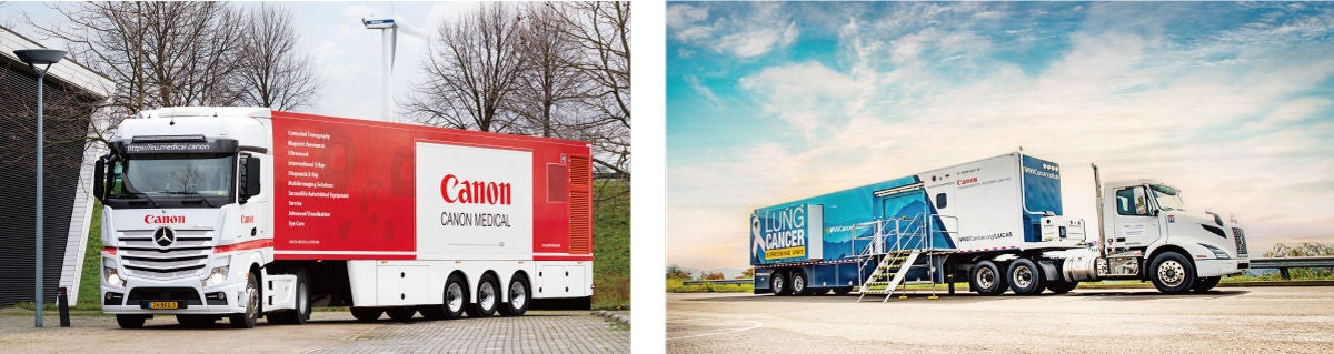
One of the main challenges of lung cancer screening is reaching the people who may need it the most. Mobile imaging solutions allow you to provide healthcare and education where people live and work, and offer screening close to their homes. By removing barriers to access, mobile imaging services may play a significant role in improving lung cancer screening rates and contribute to more equitable healthcare. The design of our mobile medical equipment provides a comfortable and convenient environment for high-patient throughput lung cancer screening without any compromises on patient safety, workflow or image quality.
Efficient workflow
Scanning is easier than ever before
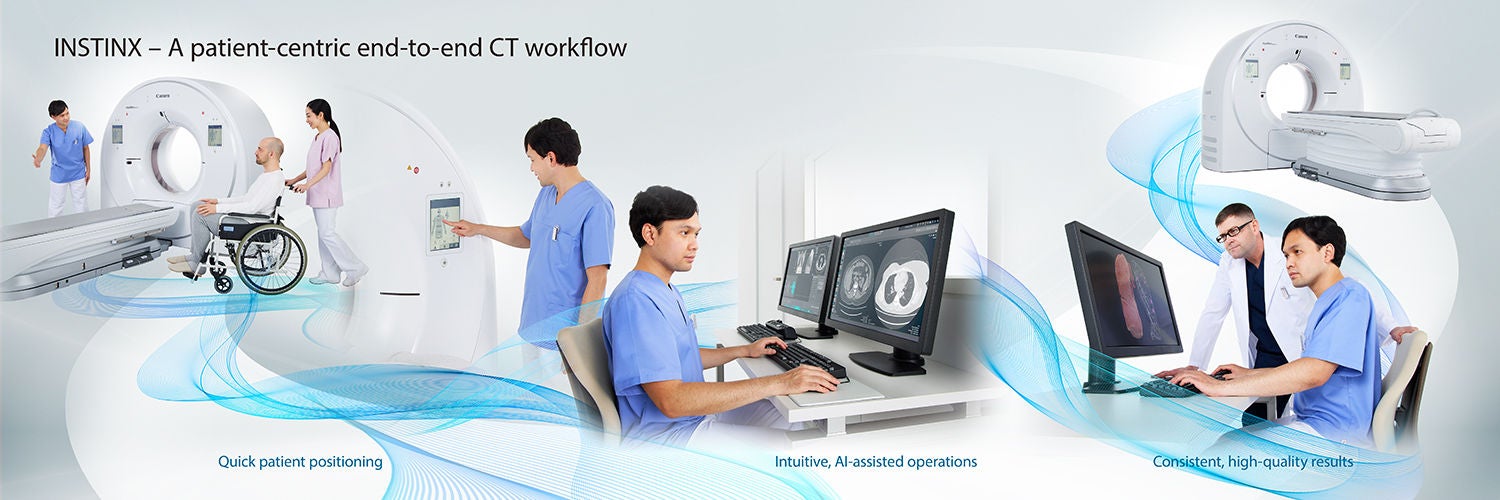
An efficient workflow is essential for lung cancer screening. It enhances accessibility, reduces waiting times, optimizes resource utilization, and improves overall patient care. By prioritizing efficiency, we provide patient-centric end-to-end workflow solutions, incorporating intuitive design and automated features for improved efficiency through the INSTINX workflow.
INSTINX workflow
Canon's CT total workflow experience is redesigned from the ground up to set new standards in efficiency and consistency. Every detail of the workflow has been thoroughly refined and validated through clinical testing in medical centers around the world.
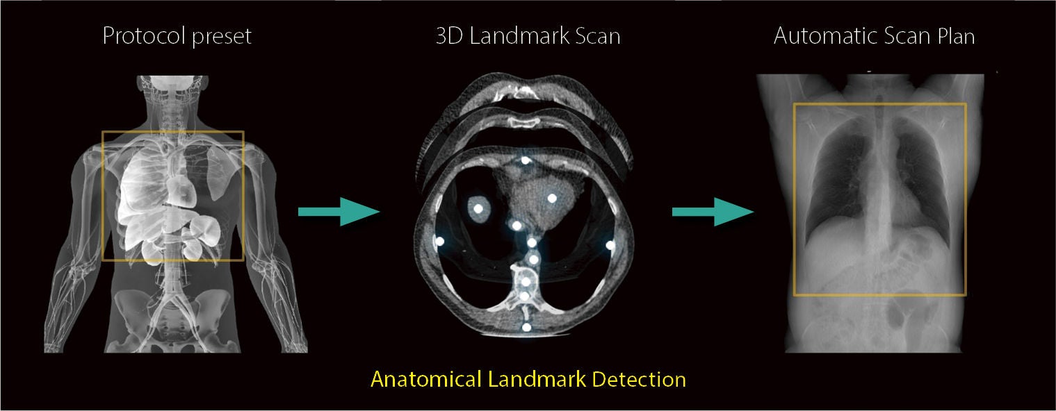
Automatic scan planning for all chest routine scans
INSTINX features automatic scan planning to automatically plan all routine scans including scan ranges and parameters, including dose and exposure settings, using the unique 3D Landmark Scan data and Anatomical Landmark Detection technology.
High-quality images at low dose
A unique synergy of beam filtering and AI-enhanced reconstruction technologies
Lung cancer screening demands high patient throughput and low radiation dose while providing images that have the quality for a reliable assessment of nodule growth and malignancy. The unique combination of SilverBeam Filter with our specific Deep Learning imaging reconstruction results in the best possible image quality at typical lung cancer screening doses.

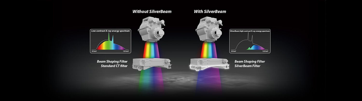
SilverBeam Filter
SilverBeam, a beam shaping energy filter, leverages the photon-attenuating properties of silver to selectively remove low energy photons from a polychromatic X-ray beam, leaving an energy spectrum optimized for lung cancer screening.
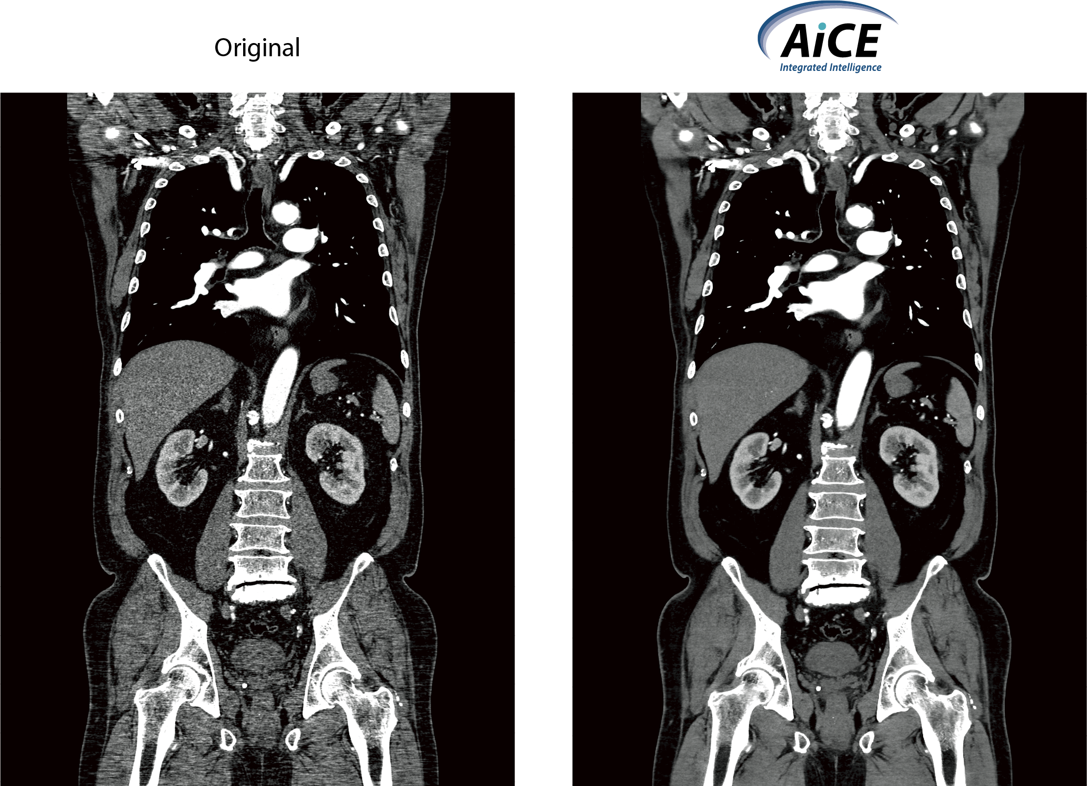
Advanced intelligent Clear-IQ Engine (AiCE)
AiCE is an innovative Deep Learning Reconstruction technology that has been trained to reduce noise and boost signal to deliver sharp, clear and distinct images at speed.

Small lung nodule detection
High-quality lung CT imaging at 0.54 mSv utilizing the combination of SilverBeam Filter and AiCE.
In this patient, a small lung nodule is clearly visible in the upper right lobe.
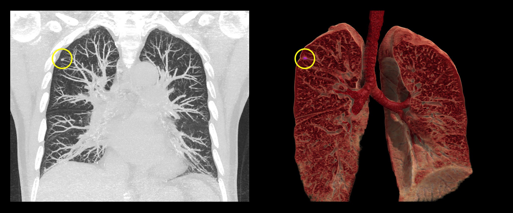 Courtesy of Fujita Health University Hospital, Japan
More detail in comparison to X-ray
In contrast to the chest X-ray, the presence of lung cancer is clearly visible in the left upper lobe. Utilizing the combination of SilverBeam Filter and AiCE, the effective dose of this scan was only 0.18 mSv, a dose level closer to a chest X-ray3
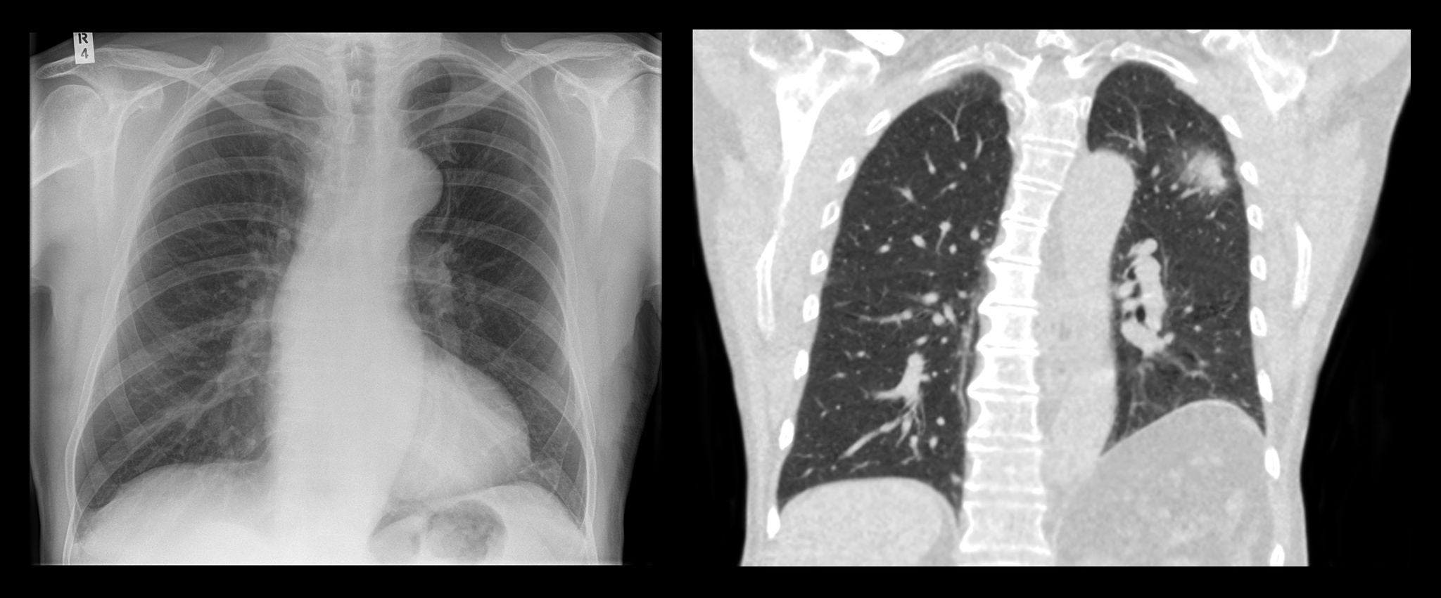 Courtesy of Dr. Russell Bull, Royal Bournemouth Hospital, Bournemouth, UK
Follow-up imaging at low dose
SilverBeam Filter combined with AiCE delivers high-quality images of the lungs at a dose closer to a chest X-ray.
In this patient, 94% dose reduction was achieved at follow-up through the implementation of our SilverBeam Filter as compared to the initial scan using a standard filter, while nodule conspicuity was maintained.
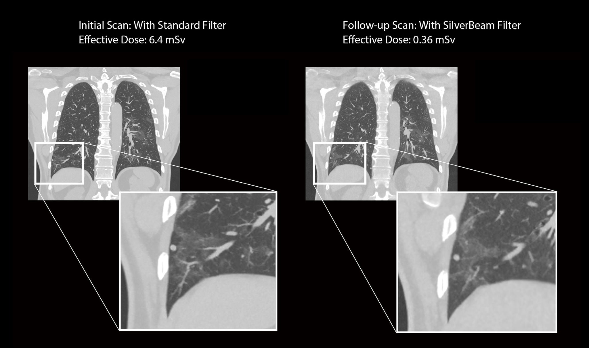 Courtesy NHLBI, National Institutes of Health, USA
Risk stratification and diagnosis
Standardized lung nodule assessment and comprehensive characterization of nodules over time
A key aspect of any lung cancer screening program is the accurate and timely reporting of scans followed by streamlined nodule management and lung disease management. However, without appropriate software solutions, the reporting of lung cancer examinations can be a time- consuming task that is prone to error.
The comprehensive lung cancer screening solution in Vitrea Advanced Visualization contains automated tools that support you in the detection and characterization of different lung nodule types including the assessment of growth patterns. To facilitate workflow and structured reporting, findings are automatically transferred to your reporting template with quick reference to guidelines such as Lung-RADS and the Fleischner Criteria.
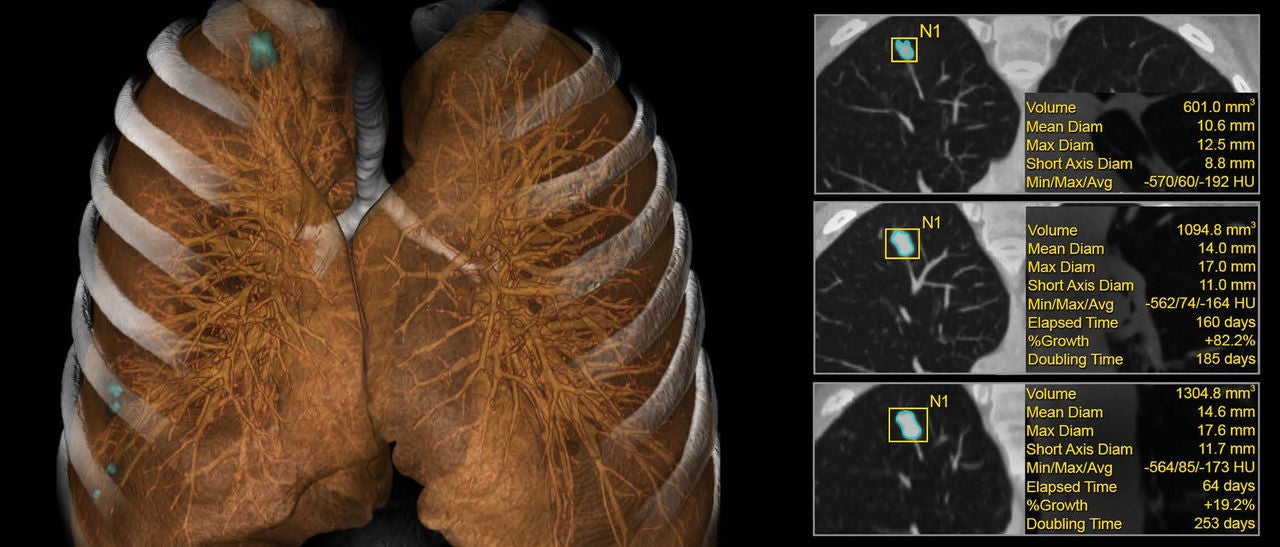 Automatically detects potentially actionable lung nodules and quantitative assessment of growth.
Clinical Evidence
Customer Presentations
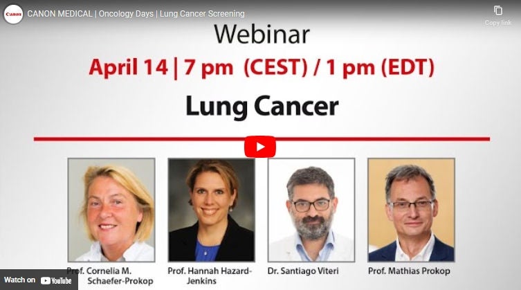
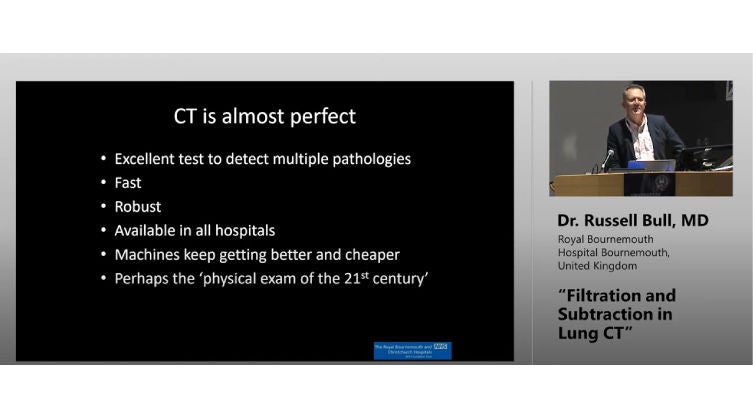
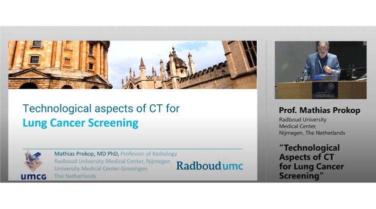
Watch our speakers present the latest developments in lung cancer screening, give advice on how to overcome barriers in clinical implementation and improve access to screening and share the newest technology in ultra-low dose CT.
White Papers and Case Studies
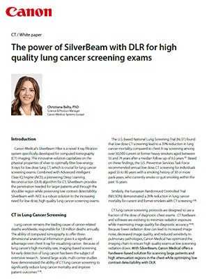
Download our latest white papers and case studies on lung cancer screening solutions using Canon technologies here.
Scientific Papers
Find our latest scientific evidence on lung cancer screening solutions using Canon technologies here.
Yanagawa, M et al. | Lung adenocarcinoma at CT with 0.25- mm section thickness and a 2048 matrix: high-spatial-resolution imaging for predicting invasiveness |Radiology (2020)
Tsubamoto, M et al. | Ultra high-resolution computed tomography with 1024-matrix: Comparison with 512-matrix for the evaluation of pulmonary nodules. |European Journal of Radiology (2020)
Lucia J M Kroft et al. | Added Value of Ultra-Low-Dose Computed Tomography, Dose Equivalent to Chest x-Ray Radiography, for Diagnosing Chest Pathology | J Thorac Imaging (2019)
Fujita, M et al. | Lung cancer screening with ultra-low dose CT using full iterative reconstruction | Japanese journal of radiology (2017)
Meyer, E. et al. | Wide-volume versus helical acquisition in unenhanced chest CT: prospective intra-patient comparison of diagnostic accuracy and radiation dose in an ultra- low-dose setting |European Radiology (2019)
Schaal, M. et al. | Diagnostic Performance of Ultra-Low-Dose Computed Tomography for Detecting Asbestos-Related Pleuropulmonary Diseases: Prospective Study in a Screening Setting| PLOS One (2016)
Kakinuma R et al. | Ultra-High-Resolution Computed Tomography of the Lung: Image Quality of a Prototype Scanner | PLoS ONE (2015)
Nomura, Y et al. | Effects of iterative reconstruction algorithms on computer-assisted detection (CAD) software for lung nodules in ultra- low- dose CT for lung cancer screening |Academic Radiology (2017)
General references
3. RadiologyInfo.org | Radiation Dose from X-Ray and CT Exams | RadiologyInfo.org. (2022)
Social Media