
Dynamic Volume CT
MSK Imaging
True 4D CT made possible.
Faster, More Accurate Diagnosis
Superior treatment decisions for MSK made possible
Canon Medical’s road to dynamic volume CT began decades ago. In 2007, we introduced the Aquilion ONE Area Detector CT. Next to routine anatomical imaging, it provided access to high-resolution isotropic 160mm volume datasets to visualise joint movement or any other 4D dynamic process. Dynamic volume scanning is able to capture the often-complex mechanical abnormalities causing the patients symptoms that can not be visualised with other imaging techniques.
As such, superior treatment decisions can be made for a wide variety of musculoskeletal functional disorders.
Contact your Canon Medical representative to start using Dynamic Volume CT on your Aquilion ONE scanner.
Capturing motion during imaging
Dynamic volume CT scans are just like a normal CT scan with the exception being that the patient will be asked to perform movements during the scan.
Dynamic volume CT scans will provide the clinician with unique 4D information about joint motion. The high resolution 4D information provided by these scans is not available using other techniques, including X-ray, Ultrasound and MRI.
Standard CT wrist scan reconstructed as a static 3D image. Provides good detail about bone position and structure (left). Dynamic volume CT scan of wrist reconstructed as a dynamic 4D movie provides good detail about bone position and structure as well as enabling assessment of complex joint motion (right).
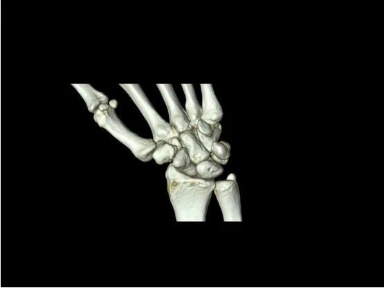
With a wide area detector covering 16 cm, Aquilion ONE CT systems can capture entire joints within one rotation and subsequently provide 4D data sets of orthopaedic joints and complex motions that can be viewed over time.
1. Wide Area Detector
With a wide area detector covering 16cm in a single rotation with every slice just 0.5mm thick...
2. Dynamic Joint Motion
We are able to capture complex joint motion...
3. 4D CT
and display that information in an interactive high resolution 4D CT image.
Dynamic Volume CT MSK Imaging
True 4D CT MSK imaging made possible
With Canon Medical's dynamic volume CT technology, true 4D imaging can be obtained of selected joints within the body including the wrist, elbow, ankle, knee, shoulder, S/C joints and A/C joints.
Below are a few examples.
Dynamic Volume CT Imaging – Shoulder
This 4D Dynamic Volume CT performed on the Aquilion ONE demonstrates the dislocation of the AC Joint in both the 3D Volume Render (Right) and Coronal MPR (Left). The pathology was best visualised by having the patient replicate a movement that caused them severe discomfort, in this case putting on a car seatbelt.
Courtesy of Victoria House Medical Imaging Melbourne
Dynamic Volume CT Imaging - Ribs
Dynamic Volume CT Imaging - Elbow
Dynamic volume elbow CT performed to assess range of motion, prior surgery to biceps tendon insertion. Patient was experiencing pain on radial ulnar deviation.
Courtesy Flinders Medical Centre, Adelaide, Australia
Dynamic Volume CT Imaging - Patella
Dynamic volume patella CT scan to assess patella maltracking.
Courtesy Monash Medical Centre, Melbourne, Australia
Post-process your Dynamic Volume CT for better image quality and reporting
4D Orthopaedic Software
Customer story | Victoria House Medical Imaging (VHMI)
Achieving CT MSK imaging excellence with dynamic volume scanning
Victoria House Medical Imaging (VHMI) is recognised as the premier provider of musculoskeletal and sports imaging services in Victoria and has been in practice for almost 30 years. Watch the customer story to find out how Dynamic Volume 4D CT imaging with the Aquilion ONE has helped them to address the challenges they have been facing and improve the confidence in diagnosis.
Dynamic Volume CT Imaging
Patient Instruction Videos
Depending on the examinations, a patient will be required to perform one or more movements during the scan. Watch the instruction videos to find out how to get your patients prepared for a Dynamic Volume CT scan.

Social Media